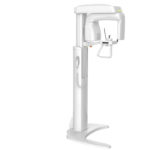
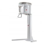
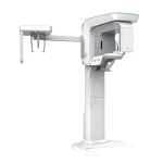
Cypher E is a new generation digital panoramic X-ray unit providing clear images with excellent operability.
Cypher E is capable of smooth exposure without requiring the patient in an uncomfortable posture, thus when aligning the positioning beams to a patient, Cypher E moves to the position automatically with the beams.
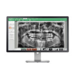
The X-ray image is displayed on a PC monitor in real time. The versatile image processing function of the software provides the diagnosis information necessary to provide advanced dental care and treatment, and supports the clinic to enhance the quality of informed consent.
Full Digital
- Easy conditional setting for exposure
- Reduction of X-ray dosage
- IT system for dental clinic
- Enhancement of quality of informed consent
- Environmental awareness
Panoramic exposure: 10 seconds
Tomosynthesis
Acquisition of Panoramic images in Tomosynthesis mode provides image data with a slice depth of 30mm.
It is now possible to clearly see the blurring of the anterior teeth image area even in positioning failure.
*For children, the acquisition area of panoramic image data is different.
*This function is available only in NEOPREMIUM2.
- Automatic display can be performed with optimal slice positioning for the anterior teeth, from a region with a slice depth of 30 mm.
- It is further possible to select an image from each of the anterior teeth and the left and right molars to obtain a set of images best matching the shape of the patient’s dentition (for children, the acquisition area of panoramic image data is different).
- Clearer images can be displayed using data from 31 images spaced at 1 mm intervals.